


NEWS
Oct 1, 2015
Release of the Trinias series MiX package,
an Angiography System Supporting Minimally Invasive Treatments
Through Enhanced Applications
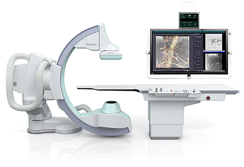
Trinias F12 MiX package Floor-Mounted C-Arm Type
Shimadzu has released the Trinias series MiX package, an angiography system providing enhanced therapeutic support applications.
In comparison to invasive surgical procedures, minimally invasive procedures use a catheter (a small tube for medical procedures), which is inserted into a blood vessel through a small incision in the skin. X-ray fluoroscopy is used during the procedure to guide the catheter and deploy devices. Endovascular therapy is less invasive and more patient friendly and as a result, widely used in the context of heightened consciousness of Quality of Life (QOL).
Angiography systems must provide high imaging quality and ease of operation to support the practitioner during procedures.
It is expected that advances in stents and other endovascular therapeutic devices in recent years, as well as the establishment of various interventional procedures will further reduce the burden on patients.
Shimadzu has extended the Trinias angiography system, which facilitated high-level interventions through our proprietary image processing technology, and has developed the "Minimally invasive eXperience (MiX)" package to support less invasive treatments through a variety of applications. The newly developed 3D application allows the angiography system to be linked to previously acquired CT images during the interventional procedure. The trace mapping function automatically extracts and displays only the outline of vessel walls providing a map image for easy device guidance.
In addition, the PCI (Percutaneous Coronary Intervention) support application can display stents in a fixed position in real time. As a result of these functional enhancements, shorter treatment times are achieved and less contrast media is used.
Furthermore, thanks to the SCORE PRO Advance image-processing engine, the system now can operate at about half the conventional X-ray dose. Together, these support more user-friendly treatment, with less burden on patients.
Features of the New Package
Enhanced Therapeutic Support Applications
1. SCORE Navi + Plus 3D Application Interlinked with CT Images
With this application, you can import pre-procedure CT images to the workstation, enabling an overlay with fluoroscopy, including angular synchronization with the angiography system C-arm. Linking the CT images directly to the treatment images allows seamless flow from pre-procedural therapeutic design to navigation during the procedure.
2. SCORE StentView + Plus PCI Support Application Improves Device Detection Efficiency
This is the more advanced version of Shimadzu's propriety SCORE StentView application to support safe and reliable PCI. In addition to a function enabling stents, which are in constant motion due to heartbeats, to be enhanced and displayed in a fixed position in real time, a new function has been added to configure the region of interest (ROI) from the preceding fluoroscopy image. Narrowing the detection area on the fluoroscopy image dramatically improves the device detection efficiency, even in procedures using multiple devices, thereby shortening treatment times.
3. SCORE MAP Provides Automatic Trace Mapping, Useful for Aortic Stent Grafting
This newly developed trace mapping function automatically extracts only the outline of vascular walls based on DSA* images. Overlaying this with fluoroscopy images dramatically increases the visibility of wires and devices. This will be useful in endovascular treatments in the aorta, where contrast concentration uniformity is particularly problematic, while helping to reduce contrast media use. In addition, the mapping region can be narrowed and sketched by the practitioners themselves, supporting more efficient procedures.
* DSA (Digital Subtraction Angiography):
This is a radiography procedure in which images of bone and blood vessels taken after the administration of a contrast media are processed to digitally subtract the information on the bones only in real time, thereby extracting just the blood vessels.
For more details, visit here.

- FOLLOW US ON SOCIAL MEDIA


